For Patients
Want to know where this treatment is offered?
Find a Clinic
Frenectomy treatment with the laser is effectively completed in a single procedure for optimal results.

Recovery is minimal, with most patients resuming normal activities within 24-48 hours after the procedure.

The treated area heals smoothly, leaving little to no visible scarring.

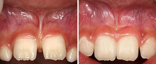
BEFORE
AFTER
Our treatments and systems are trusted throughout the industry thanks to our commitment to quality, durability, and innovation.

LIFE-CHANGING OUTCOMES
Our devices are recognized for their exceptional performance, offering patients minimally invasive, comfortable treatments for transformative results.
product excellence
Our dedication to perfection ensures each product delivers superior treatments patients can trust.
industry leaders
Our ongoing research enables us to push the limits of medical technology and set new standards of excellence among our peers in a rapidly changing field.
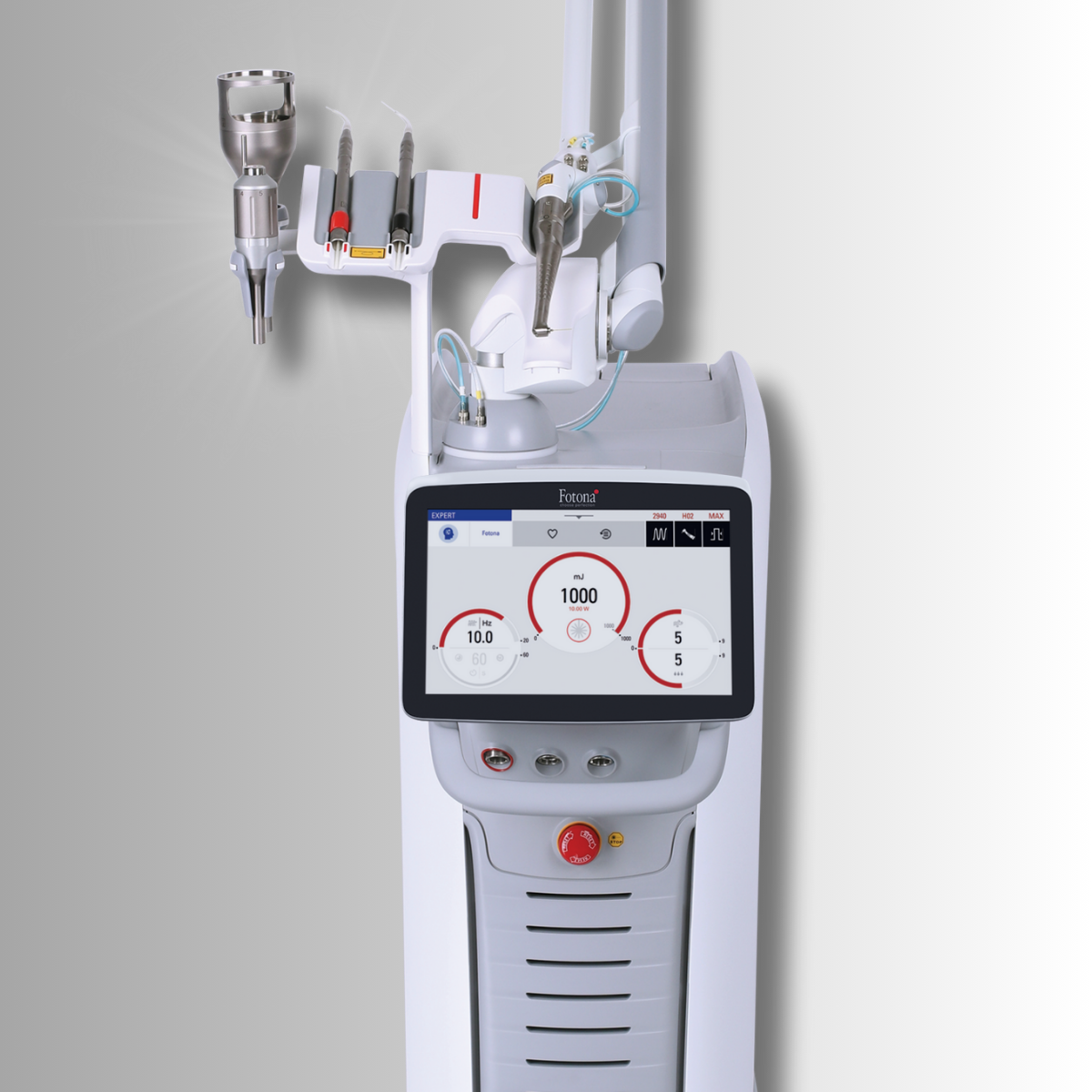
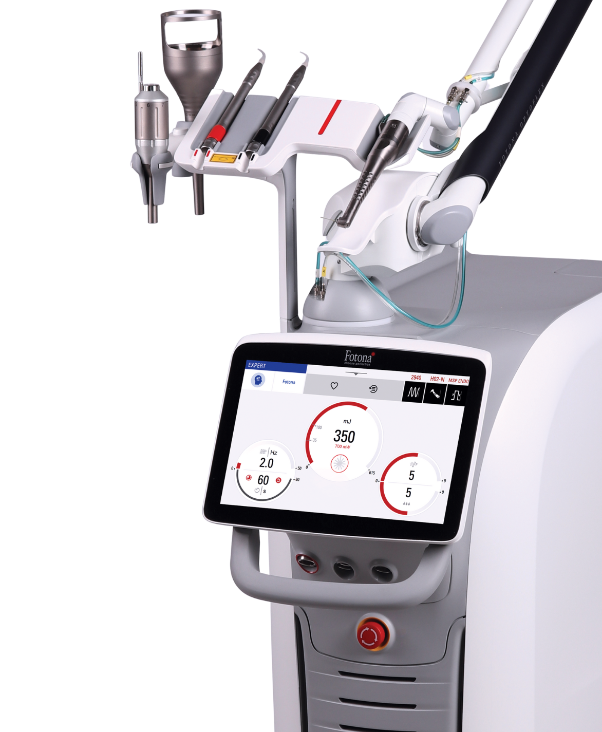
Powerful Dual-Wavelength Dentistry Laser with Sleep and Aesthetic Possibilities
Frequently Asked Questions
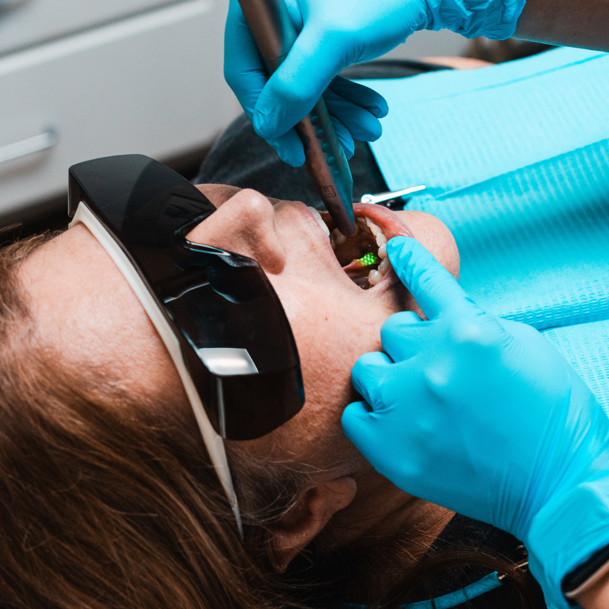
Frenectomy Treatment
Expand the capabilities of your laser with accessories built to adapt with your evolving practice.
No Accessories Found
This presentation will summarize and present video of some of the most common procedures performed day-to-day with the Fotona LightWalker® ATS and its dualwavelength capability.
In these three case reports we describe the use of Nd:YAG laser (AT Fidelis, Fotona d.d. Ljubljana, Slovenia) on correcting frenulum pull and increasing the keratinized mucosa around affected teeth using a frenectomy and vestibuloplasty procedure.
In this clinical case, a bonded RPE was placed in a 10-year-old male patient, for class Ill unilateral posterior and anterior crossbite correction. (Figure 1). Anterior alignment was achieved by bracketing upper 2-2 with the NiTi wire engaging bracket attachments on the maxillary RPE in the maxillary 4 locations. Expansion was maintained with composite on the expansion screw for 2-3 months after full expansion (Figure 2).